Scientists believe they may have found a way to detect early signs of a rare form of dementia using a simple eye test.
Researchers from the University of Pennsylvania School of Medicine say that signs of frontotemporal dementia (also known as frontotemporal lobe degeneration or FTD) can be seen in the retina.
FTD is an uncommon type of dementia that is hard to diagnose, but the scientists have found that thinning of the outer retina could indicate the person may have the disease.
According to Alzheimers Research UK, FTD is believed to account for less than 5% of all dementia cases.


It is caused by damage to brain cells in the frontal lobe, which regulates our emotions, behaviour and reasoning, and the temporal lobe – which involves the understanding and production of language.
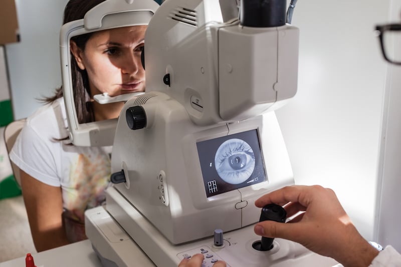
The researchers now say that a simple eye exam and retinal imaging test can detect a thinning of the outer retina – and previous studies have suggested that other neurodegenerative diseases, like Alzheimer’s and motor neurone disease, can also cause a different part of the retina to thin.
Lead author Dr Benjamin Kim, assistant professor of Ophthalmology, said: “Our finding of outer retina thinning in this carefully designed study suggests that specific brain pathologies may be mirrored by specific retinal abnormalities.”
The scientists studied 38 FTD patients and 44 control subjects who did not have any neurodegenerative disease and used an eye-imaging technology called spectral-domain optical coherence tomography (SD-OCT) to measure retinal layers.

The SD-OCT is non-invasive and uses a safe light beam to image tissue with micron-level resolution.
Measurements revealed that the outer retinas of the FTD patients were thinner compared to the control subjects, which was caused by a thinning of two specific portions of the outer retina – the outer nuclear layer (ONL) and ellipsoid zone (EZ).
The ONL of FTD patients was about 10% thinner than controls and was the primary source of the outer retina thinning.
The results suggested FTD manifests in a different way in the structures of the retina and detecting this difference might help doctors distinguish one disorder from another.
The study is published in Neurology.